dna/rna recognition by natural and artificial binders
Large variety of small molecules and supramolecules, including biomimetics, have been investigated, over the last few decades, as agents for DNA/RNA recognition and binding, with an aim of the development of novel (anticancer) drugs, biosensors or tools for basic biochemical and biological studies.1-3 Unique architecture of nucleic acids, characterized by a combination of an anionic, electron-rich phosphate backbone, abundance of electron-donor heteroatoms from the nucleobases, as well as by sophisticated secondary (and tertiary) structures, create an attractive and very complex target for many external recognition agents including metal complexes. There are two essential modes of interaction with a nucleic acid chain: covalent and non-covalent. While the first involves an adduct formation and an irreversible modification of a target site, the latter is rather reversible and, apparently, less invasive. The specific way in which particular molecule will recognize a precise sequence within the DNA or RNA chain depends on its structural and physico-chemical properties. For instance, there are four major ways of interaction with the double stranded DNA chain: intercalation between hydrogen bonded nucleic pairs of a double helix, electrostatic interactions with the electron-rich phosphate backbone, minor groove interactions and major groove interactions. To mention only that recently, versatile molecules capable of combining two or more of these recognition modes were more thoroughly investigated.4 Importantly, the two latter zones of interaction (major and minor groove) are favourable for the molecular agents with a task to target specific genes, as within the grooves, the base sequence is widely exposed and easily readable. Specific chemical and sterical environment in the major groove, featuring large variety of H-bond donors and acceptors, makes it an ideal molecular target within the double stranded DNA structure.5 Major grove is, therefore, a preferred site for DNA recognition which is extensively used by Nature itself, as its dimensions are perfectly suited to accommodate variety of typical protein motifs.6 Electrostatic interactions and coordinative binding are generally accepted modes of interaction of bonding molecules with all types of nucleic acids. The association of binding molecules (artificial or natural) and the DNA/RNA targets modify their physico-chemical and structural characteristics and elicit new properties, which makes this process of interest for applications in many different technologies the most outstanding of which are drug design and development of biosensors to be used in bioanalytics and diagnostics. In contrast to proteins, non-proteic ligands generally are small molecules that interact either with the minor groove7 or undergo intercalation (polyaromatics for instance) in between paired bases. Surprisingly, the number of such, artificial ligands that specifically interact in the major groove is very small comparatively. Only relatively recently, the first artificial (non-covalent) major groove binders have been reported by Hannon and co-workers.8 Interaction of (biomimetic) artificial binders with the major groove, therefore, is a large, unexplored area of a potentially exciting research with an enormous applicative potential.
Structural research of nucleic acids
To recognise the (non-covalent) interactions responsible for the formation of macromolecular complexes (and hence for the new properties generated by the process of association of binding molecules), it is essential to determine the 3D structures of the macromolecular complexes formed by these interactions. Only then becomes it possible to precisely correlate the structural features of the binding molecules with a newly generated properties, and hence to inspire the design of the novel generations of binders which would be able to specifically tune the desired properties of the targeted macromolecules. Single crystal X-ray diffraction is, undoubtly, a method of choice in elucidating the details of the molecular structures of nucleic acids, which are, in the X-ray diffraction studies represented by the specially prepared oligonucleotide chains.9 However, as production of good quality single crystals of oligonucleotides, due to their pronounced conformational flexibility, has historically been a notorious challenge, NMR has been imposed as an alternative method of nucleic acid structure elucidation.10 This method has an additional advantage that the structures are solved in solution (which may sound more physiologically relevant). At the same time, however, NMR as a method in elucidating oligonucleotide structures has serious limitations, such as the dimensions of the oligonucleotide chain that can possibly be structurally characterized, as well as the fact that reliable structural information can be deduced only for specific portions of the structure. Finally, molecular modelling techniques enable calculation and visualization of dynamic changes of the structure and conformation, and, as such, provide an invaluable additional, complementary set of information on the structural features of a given macromolecule to the single crystal X-ray diffraction and NMR.
Calixarenes/resorcinarenes as dna/rna binders
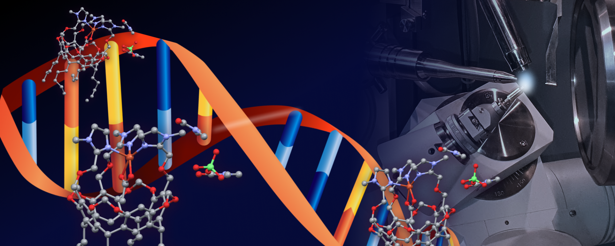
The research on calixarenes as preorganized platforms for the binding of metal ions and small neutral molecules has been popular for over three decades.11,12 Calixarenes are remarkable supramolecules well known for their ability to bind to many biological molecules including nucleic acids fragments and DNA/RNA polymers.13 Importantly, calixarenes generally easily penetrate into the cell and are stored in the cytoplasm.14 These calixarene features are of significance for various (biological) applications based on nucleic acids recognition. The versatile nature of calixarenes and possibility of plethora of their chemical modifications including size, conformation, stereochemical availability of binding groups and amphiphilicity15 have been an inspiration for researchers to probe variety of their applications, spanning from life sciences16 all the way to molecular sensors research and development of smart materials.17 There is one more advantage of their easy chemical modifications; the active small groups (e. g. fluorophores) can be easily grafted to the calixarene/resorcinarene scaffold, in order to elicit spectroscopic signal(s) for an easy detection of newly formed biocomplexes. The ability of these remarkable molecular edifices to bind the DNA chain was reported recently by us (Figure A1).18

There are a few 3D structures of protein-calixarene assemblies determined and deposited in Protein Data Bank, whereas absolutely no X-ray data is available, at this point, on 3D molecular architectures of DNA/RNA – fragments bound to calixarene- or resorcinarene-based binders.19 It is, therefore, a prodigious challenge for us to explore binding of our, suitably modified, biomimetic calix[6]arene-based (funnel) and resorcinarene-based “bowl” complexes20 to nucleic acids, and finally characterize chemically and structurally the emerged biocomplexes.
REFERENCES
- I. Renard, M. Grandmougin, A. Roux, S. Y. Yang, P. Lejault, M. Pirrotta, J. M. Y. Wong, D. Monchaud, Nucleic Acids Research, 47 (2019) 5502 – 5510.
- N. Mohajeri, M. Imani, A. Akbarzadeh, A. Sadighi, N. Zarghami, Biosensors and Bioelectronics, 144 (2019) 111633.
- M. Giuliani, I. Morbioli, F. Sansone, A. Casnati, Chem. Commun. 51 (2015) 14140 – 14159 and referencese herein.
- B. Willis, D. P. Arya, Biochemistry, 49 (2010) 452 – 469.
- P. L. Hamilton, D. P. Arya, Nat. Prod. Rep. 29 (2012) 134 – 143.
- S. Neidle, Nucleic Acid Structure and Function, Oxford University Press, Oxford, 2004.
- L. Steiningerova, Z. Kamenik, R. Gazak, S. Kadlcik, G. Bashiri, P. Man, M. Kuzma, M. Pavlikova, J. Janata, J. Am. Chem. Soc. 2020, DOI: 10.1021/jacs.9b11234.
- (a) M. J.Hannon,V.Moreno,M. J.Prieto, E. Moldrheim, E. Sletten, I. Meistermann, C. J. Isaac, K. J. Sanders, A. Rodger, Angew. Chem. 113 (2001) 903–908; (b) M. J. Hannon, V. Moreno, M. J. Prieto, E. Moldrheim, E. Sletten, I. Meistermann, C. J. Isaac, K. J. Sanders, A. Rodger,Angew. Chem. Int. Ed. 40 (2001) 879– 884; (c) I. Meistermann, V. Moreno, M. J. Prieto, E. Moldrheim, E. Slatten, S. Khalid, P. M. Rodger, J. C. Peberdy, C. J. Isaac, A. Rodger, M. J. Hannon, Proc. Natl. Acad. Sci. USA 99 (2002) 5069–5077.
- B. Rupp: Biomolecular crystallography; Principles, practice and application to structural biology, Garland Science, Taylor and Francis Group, New York, USA, 2010.
- (a) T. L. James (Ed.), (2005) Nuclear magnetic resonance of biological macromolecules. Methods in Enzymology, Volume 394. (b) M. P. Latham, D. J. Brown, A. McCallum, A Pardi, Chembiochem. 6 (2005). 1492–1505; (c) S. D. Kennedy (2016) NMR Methods for Characterization of RNA Secondary Structure. In: Turner D., Mathews D. (eds) RNA Structure Determination. Methods in Molecular Biology, vol 1490. Humana Press, New York, NY.
- L. Baldini, F. Sansone, A. Casnati, Cation Complexation by Calixarenes, Elsevier Reference Module in Chemistry, Molecular Sciences and Chemical Engineering, Elsevier, Waltham, MA, 2013, DOI: 10.1016/B978-0-12-409547-2.05616-X.
- L. Baldini, F. Sansone, A. Casnati, R. Ungaro, Calixarenes in molecular recognition, in Supramolecular Chemistry: from Molecules to Nanomaterials, ed. J. W. Steed and P. A. Gale, John Wiley & sons, Chichester, 2012, vol. 3, pp. 863–894.
- (a) Peters, M. S., Li, M. & Schrader, T. Nat Prod Commun 7 (2012) 409–417; (b) Shi, Y. H. & Schneider, H.-J. J. Chem. Soc. Perkin Trans. 2 (1999) 1797–1803.
- R. Lalor, H. Baillie-Johnson, C. Redshaw, S. E. Matthews, A. Mueller, J Am Chem Soc. 130 (2008) 2892 – 2893.
- Han-Wen Tian, Yan-Cen Liu, Dong-Sheng Guo, Mater. Chem. Front. 4 (2020) 46.
- (a) F. Sansone, L. Baldini , A. Casnati, R. Ungaro, New J. Chem., 34 (2010) 2715-2728; (b) V. Rullaud, N. Moridi, P. Shahgaldian, Langmuir, 30 (2014) 8675-8679; (c) R. Rodik, S. Cherenok, O. Kalchenko, O. Yesypenko, J. Lipkowski, V. Kalchenko, Current Organic Chemistry, 22 (2018) 2200 – 2222; (d) Lin An, Jia-wei Wang, Jia-dong Liu, Zi-ming Zhao, Yuan-jian Song, Frontiers in Chemistry (2019) doi.org/10.3389/fchem.2019.00732; (e) S. Engilberge, M.L. Rennie, E. Dumont, P.B. Crowley, ACS Nano. 13 (2019) 10343-10350.
- R. Kumar, A. Sharma, H. Singh, P. Suating, H. S. Kim, K. Sunwoo, I. Shim, B. C. Gibb, J. S. Kim, Chem. Rev. (2019) DOI: 10.1021/acs.chemrev.8b00605.
- W. Hu, C. Blecking, M. Kralj, L. Šuman, I. Piantanida, T. Schrader, Chemistry, Eur. J., 18 (2012), 3589–3597.
- To access Protein Databank free of charge, go to https://www.ebi.ac.uk/pdbe/node/1
- J.-N. Rebilly, B. Colasson, O. Bistri, D. Over, O. Reinaud, Chem. Soc. Rev. 44 (2015)467-489. and references therein.
